|
shy
Crypt Keeper
 
Australia
1991 Posts |
 Posted - 26 March 2005 : 9:09:45 PM 


|
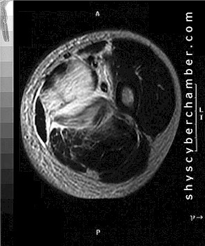
Contributor's Note:
Myositis ossificans is a benign, ossifying soft-tissue lesion typically occurring within skeletal muscle. Patients are usually adolescents and young adults; myositis ossificans is rare in children under 10 years of age. The most frequent symptoms and signs are pain and tenderness with a soft tissue mass. Approximately 80% of cases arise in the large muscles of the extremities.
Grossly, the lesion tends to be well circumscribed. On cut surface, it is white, soft, and rather gelatinous (or hemorrhagic) in the center, and yellow-gray and firm with a rough granular surface at the periphery.
Microscopically, myositis ossificans is characterized by a distinct zonal pattern. This pattern is most conspicuous in lesions of three or more weeks' duration. There are three distinct zones: a central portion in which there is fibroblastic proliferation which can vary markedly in cellularity, pleomorphic characteristics, and numbers of mitotic figures; an intermediate portion where there is collagen and osteoid deposition among proliferating spindle cells, with early trabeculation of ossifying areas; and a peripheral zone of osteoid trabeculae rimmed by osteoblasts, with bone surrounded by loose fibrous tissue and atrophic fat. Characteristically, in myositis ossificans the bone formation is most prominent at the periphery of the lesion.
The differential diagnosis of myositis ossificans includes other extraskeletal bone-forming lesions such as fibrodysplasia (myositis) ossificans progressiva and osteosarcoma. Fibrodysplasia (myositis) ossificans progressiva is a rare, slowly progressive hereditary disease that principally affects children under the age of ten years. It is associated with symmetrical malformations of the digits (especially microdactyly) and other skeletal malformations. The disease is characterized by progressive fibroblastic proliferation and subsequent calcification and ossification of subcutaneous fat, muscles, tendons, aponeuroses, and ligaments. Unlike localized myositis ossificans, the ossification occurs in the center of the nodules.
Differentiating between myositis ossificans and extraskeletal osteosarcoma is critical. The zoning pattern of growth in myositis ossificans is an important diagnostic point. Osteosarcoma displays disorderly growth of atypical hyperchromatic and pleomorphic tumor cells. Bone deposition in osteosarcoma is haphazard and disorganized, sometimes with a "reverse zoning effect", with bone formation in the center of the lesion and immature spindle cells toward the periphery. Mitotic figures are present in both the immature, central portion of myositis ossificans and osteosarcoma, but myositis ossificans lacks the atypical or tripolar mitotic figures characteristic of osteosarcoma.
Diagnostic problems may arise in myositis ossificans if a biopsy is small and taken only from the central area in the early proliferative phase, as this may lead to confusion with osteosarcoma or a soft tissue sarcoma. An adequate open biopsy will display the zoned pattern and exclude malignant entities from consideration.
Because myositis ossificans is a benign self-limiting process, the prognosis is excellent. Local excision is curative.

|
|


 Left Thigh Mass, Biopsy: Myositis Ossificans [p]


